User-agent: Google Allow: A Simple Guide to Medical Conditions: physiotherapy

Search This Blog

Showing posts with label physiotherapy. Show all posts

Sunday, December 18, 2011

PHYSIOTHERAPY

DOC WHAT IS PHYSIOTHERAPY

Physiotherapy is also known as physical therapy of medical conditions. That answers the question of what is physiotherapy for many people.

As a supplementary type of health care, physiotherapy concerns itself with providing physical healing methods for many different kinds of injuries and illnesses.

Therapy at a Physiotherapy Clinic:

When a patient is referred to the physiotherapy clinic, he or she will be evaluated by a physiotherapist.

After this initial evaluation, he or she will be scheduled for treatments like ultrasound or acupuncture etc as recommended by the doctors in conjunction with the physiotherapists.

They will be assigned exercises to do at the clinic. A good physiotherapist will begin treatment right away.

The different types of Physiotherapy:

1.massage or manipulation (hands-on) of the musculo-skeletal system when the muscles and tissues are injured.

2.Traction of the skeletal system to lengthen the spaces between bones so that nerves or tendons are pressed on by the bones

3.Strengthening of the muscular system helps the patient to recover from surgery as well as prevention of tightening of the muscles and tendons and making them more flexible.

4.Heat (heat pads, infra-red light, shortwave diathermy) help the blood circulating after injuries and help earlier recovery

5.ice (cold compress) help to reduce swelling and tissue damage

6.ultrasound treatment, radiofrequency waves , are all useful to relieve pain and stiffness.

7.Hyperbarbaric treatment: Increase in oxygen pressure can improve the faster healing of injuries and recovery from surgery.

All these methods tend to promote better health, both physical and psychological.

The importance of physiotherapy equipment:

Equipment for helping patients regain their strength and mobility are a part of what is physiotherapy. This equipment may allow a person who is partially paralyzed to get the most exercise possible. This is crucial in maintaining the integrity of their spines and muscles.

The importance of Education in Physiotherapy:

Besides the methods used in Physiotherapy, education is a part of what is physiotherapy. A physiotherapist will teach a patient how to care for their injuries. He will teach exercises to do at home so that therapy can continue beyond the walls of the clinic or hospital. He will teach ways to overcome difficulties that cannot be cured.

Rehabilitation is another part of Physiotherapy treatment:

Patients have injuries from sports, car accidents, or assault. These injuries can be treated through physiotherapy. Given the right treatments and an injury that will respond to treatment, much progress can be made. Full functioning may be regained. It may even be possible for them to go back to work rather than being laid up at home.

Physiotherapy is a carefully planned and executed treatment strategy.

It is based upon assessments of the conditions that patients suffer. If all goes well, the patient will return to their original condition. If this is not possible, the goal is for the patient to reach a goal that is the best movement and lack of pain that is possible.

The preventative side of the field of physiotherapy is very important in the overall holistic treatment of a patient.

It is a part of the work of practitioners of physiotherapy to encourage exercises and postures that will help patients avoid physical injuries and conditions requiring their services.

An excellent physiotherapist will have fewer return patients, but the flow of people needing physiotherapy continues.
Saturday, July 26, 2008

A Simple Guide to Plantar Fascilitis

A Simple Guide to Plantar Fascilitis
----------------------------------------------------

What is Plantar Fascilitis?
-----------------------------------------

Plantar Fascilitis (also known as Painful Heel Syndrome) is a inflammation of the plantar fascia (which stretch from the calcaneum to the toes) characterised by the pain in the heel especially in the morning and weight bearing exrcises.

It is more common in women.


What are the cause of Plantar Fascilitis?
-----------------------------------------------

The cause of plantar Fascilitis is the non-specific inflammation of the plantar fascia as a result of repetitive injury to the fascia.

In some cases the plantar fasilitis occurs as a result of a calcaneal spur impinging on the fascia.

Both heels can be affected.


What are the symptoms and signs of Plantar Fascilitis?
---------------------------------------------------------------------------------

Symptoms:

1.Pain in the heel of one or both feet

2.Pain usually is worse in the morning on getting and stepping on the floor.

3.Certain weight bearing exercises like jogging or brisk walking makes the pain worse

4.Pain is described as constant and aching

5. Pain is felt most beneath the calcaneal bone but may be present in the area of the medial arch.

Signs:

1.local tenderness in the calcaneal bone area of the heel.

2. Pain is aggravated by direct pressure.

3. It can become more painful by movement which put thethe fascia under strain such as dorsiflexion of the toes or ankles.

4.Xrays of the heel usually show no abnormally. Sometimes there is calcaneal spur which may be due to traction of the muscle or fascia on the calcaneum bone.

A stress fracture may need to be ruled out in chronic cases.


What is the Treatment of Plantar Fascilitis?
----------------------------------------------------------------

Conservative treatment:
-----------------------

Initial phase:

1.rest of the heel-avoid jogging or strenous exercises

2.Cold or ice may help reduce inflammation

3.Heel cups, cushions, tapes, pads may help to reduce the pain

4.Pain killers such as NSAID(non-steroidal anti-inflammatory drugs) for pain

5.Muscle relaxant to relax muscles

6.injection of local anesthetic and long acting steroid into the tender area of the plantar fascia may help to relieve pain.

7.Usage of a short leg walking cast for a few weeks help to avoid exertion on the inflamed fascia.

8.A cushion lined night splint which hold the foot in slight dorsiflexion is helpful.

Mobilisation phase:

Physiotherapy such as traction of the fascia and heel cord, shortwave diathermy may help once there is no pain

Surgery is usually not indicated in plantar fascilitis.

Rarely surgery is used to remove the calcaneal spur and to release the plantar fascia at its attachment to the calcaneum bone.


What is the prognosis of Plantar Fascilitis?
----------------------------------------------------------

Prognosis is usually good although healing is slow and takes up to 1-2 years.


What is the prevention of Plantar Fascilitis?
-------------------------------------------------

Avoid certain weight bearing exercises like jogging or brisk walking

Use heel cups, cushions, tapes, pads in foot wear

Lose Weight

Thursday, July 24, 2008

A Simple Guide to Knee cap Dislocation

A Simple Guide to Knee cap Dislocation
----------------------------------------------------


What is Knee cap Dislocation?
---------------------------------------------------------

Knee cap Dislocation is when the knee cap (patella) moves or slides out of place. This usually occurs on the outer side of the knee.


What are the causes of Knee cap Dislocation?
----------------------------------------------------------------------

1.Dislocated knee caps most often occur in people with loose joint ligaments.

It can occur due to sudden strain on the knee ligaments causing the kneecap to protrude out of its loose ligaments.

2.Dislocation of the knee cap may also occur due to trauma.

A sudden blow to the medial part of the knee can cause the knee cap to dislocate laterally.

3.People who are prone to dislocated knee caps usually have loose ligaments with hyperflexion of the wrists or flat feet.

This condition is usually inherited and are more common in women than in men.


What are the symptoms and signs of Knee cap Dislocation?
-------------------------------------------------------------------

Symptoms:

1.Knee cap is displaced to the lateral position

2.knee swelling and effusion due to displacement of the knee cap.

3.Knee pain and tenderness is present.

4.The knee is usually held in slight flexion.

5.There is difficulty in lifting the leg

6. Patient usually walks with a limp.

Signs:

1.Tenderness and swelling of the knee

2.Knee cap is displaced to the outside or lateral part of the knee

3.In most cases the knee cap may have returned to the central position of the knee but there is still tenderness and swelling.

4.The knee cap can move excessively from right to left.(hypermobile)

How to investigate the cause of Knee cap Dislocation?
-------------------------------------------------------------

1.examination of the knee would confirm presence of the dislocated knee cap.

There is lateral displacement of the knee cap and swelling of the knee.
Movement of the knee may be painful.

2.A knee x-ray should be done to exclude any fracture especially in the case of injury or in the elderly.

A skyline view of the knee should show the shift of the patella laterally.

3.MRI of the knee can be done to see any damage in the cartilage or meniscus of the knee.


What is the Treatment of Knee cap Dislocation?
----------------------------------------------------------------

Conservative treatment:
-----------------------

1. Most cases of knee dislocation can treated by simple reduction of the dislocated knee cap.

The heel of the leg is lifted to extend the knee and flex the hip thus relaxing the quadriceps muscles(front muscles of the thigh)

Gentle pressure is exerted on the knee cap to place it to its normal position.

The knee is then immobilized for 2-3 weeks.

Quadriceps exercises are begun as soon as possible to build back your muscle strength and improve the knee's range of motion.

Drug Therapy:
-----------------------

1.Pain killers such as NSAID(non-steroidal anti-inflammatory drugs) for pain

2.Muscle relaxant to relax muscles

Surgery:
--------------

Surgery is required if:

1.the knee remains unstable

2.Injury of the knee cap cause a partial rupture of the medial retinaculum and supporting ligaments of the knee cap.
This can cause recurrent episodes of subluxation or dislocation of knee cap.

Surgery is done to stabilize the knee cap.
Reconstruction of the quadriceps extensor muscles is done to tighten the ligaments surrounding the knee cap.


What is the prognosis of Knee cap Dislocation?
----------------------------------------------------------

Prognosis using conservative methods is fair.

Recurrences are quite common.

Preventative measures such as knee guard, quadriceps exercises, avoidance of sudden turns and twisting of the knee may help.

Prognosis after surgery is usually good as there is tightening of the knee cap ligaments and the quadriceps muscles.


What is the Prevention of Knee cap Dislocation?
------------------------------------------------------------

1.proper technique when exercising.

2.Maintain strength and flexibility of the knee by exercising the quadriceps muscles.

3.Wearing a knee guard

Tuesday, July 22, 2008

A Simple Guide to Frozen Shoulder

A Simple Guide to Frozen Shoulder
----------------------------------------------------


What is Frozen Shoulder?
-----------------------------------------

Frozen Shoulder (also known as adhesive capsulitis) is a disorder of the shoulder characterised by the slow onset of pain and restriction of movement.

It tends to be chronic and full recovery may take several months.

It is more common in women and diabetes.

Incidence is about 2 in a 1000.


What are the causes of Frozen Shoulder?
-----------------------------------------------

The exact cause is unknown but several conditions has been blamed:

1.bicipital tenosynovitis- inflammation of the biceps muscles and tendon limmiting its movements

2.rotator cuff tendonitis - inflammation of the rotator cuff muscles which surrounds the shoulder with resultant adhesions and stiffness causing limitation of movement

3.reflex sympathetic dystrophy- a disturbance in the sympatheic nervous system cause pain ine the shoulder joint and hypersensitivity of the muscles surrounding the joint. There is swelling of the arm followed by atrophy of the muscles

4.trauma - injury to the joint may cause tightening of the injured muscles around the shoulder joint.

5.Surgery of the shoulder, breast and lung may also cause frozen shoulder because of the pain resulting from the movement of the shoulder and hence stiffness of the muscles.


What is the natural progression of frozen shoulder in most cases?
-----------------------------------------------------------------------

Frozen shoulder is a disabiliting disease which can last from 5 months to 4 years.

There is chronic inflammation of the muscles surrounding the joint with adhesios formed between joint and muscles causing restriction of movement of the joint.

There is also reduced fluid in the joint further restricting movement.

Stage one("freezing" or painful stage):
There is a slow onset of pain which becomes worse and stiffening of the joint occurs.

This lasts 5 weeks to 9 months.

Stage two("frozen" or adhesive stage):
There is a slow but steady improvement in pain, but the stiffness persists.

This lasts 4 -9 months.

Stage three("thawing" or recovery):
There is a gradual return to normalcy in the shoulder motion.

This lasts 5 -26 months.


What are the symptoms and signs of Frozen Shoulder?
-------------------------------------------------------------------

Symptoms:

1.Pain in the shoulder radiating down the deltoid muscle and anterior aspect of the arm

2.Pain usually is worse at night especially lying on the affected shoulder

3.Certain movements makes the pain worse

4.Pain is described as constant, dull and aching

5.complaints of stiffness of the shoulder

6.inability to wear a shirt or blouse because of restricted movements

Signs:

1.apprehensive patient who holds the arm protectively

2.Generalised tenderness of rotator cuff and biceps muscles

3.Limited shoulder movement

4.Range of muscle movement is reduced in all directions

5.Arthrogram or MRI of shoulder can be done to confirm the diagnosis and exclude a posterior shoulder dislocation..


What is the Treatment of Frozen Shoulder?
----------------------------------------------------------------

Conservative treatment:
-----------------------
Initial phase:
1.rest of the shoulder

2.moist heat

3.Pain killers such as NSAID(non-steroidal anti-inflammatory drugs) for pain

4.Muscle relaxant to relax muscles

5.injection of local anesthetic and long acting steroid into the rotator cuff muscle may help to relieve pain.

Mobilisation phase:
1.Physiotherapy such as traction, shortwave diathermy

2.gradual mobilisation and exercises to loosen the tight muscles surrounding the shoulder

Maintenance phase:
1.Continual exercises of the shoulder muscles

2.Avoidance of strain on the muscles of the shoulder

Manipulation of the frozen shoulder may be done under anaethesia to break the adhesions and restore some movement.

Surgery is usually not indicated in frozen shoulder.
Rarely surgery is used to cut the adhesions.


What is the prognosis of Frozen Shoulder?
----------------------------------------------------------

Prognosis depends on the underlying cause.

In most cases frozen shoulder may resolve itself with time

Thursday, July 17, 2008

A Simple Guide to Prolapsed Intervertebral Disc

A Simple Guide to Prolapsed Intervertebral Disc
---------------------------------------------------------------


What is Prolapsed Intervertebral Disc?
-------------------------------------------------------


Prolapsed Intervertebral Disc is the prolapse of the intervertebral disc(which is the disc between 2 vertebrae) as a result of protrusion of the nucleus pulposus out of its weakened ligamentous ring(annulus fibrosus).

It may protrude in a posterior or postero-lateral direction causing pressure to the nerve roots especially at S1, L5 and L4 vertebrae.


What are the causes of Prolapsed Intervertebral Disc?
---------------------------------------------------------------

1.Degeneration of the posterior longitudal ligaments and annulus fibrosus occurs with age resulting in the disc being pushed out between the weakened ligaments.

2.Trauma -direct injury to disc, heavy lifting, sneezing can cuse the disc to pop out of the weakened ligaments and prolapse partially or completely.

3.Spinal tumour rarely pushes the disc out of its intervertebral space


What are the symptoms and signs of Prolapsed Intervertebral Disc?
-----------------------------------------------------------------------------

Symptoms:
1.Low Backache with pain in the lumbar region

2.Sciatica or pain shooting down 1 leg

3.Pain usually follows
severe bending
lifting heavy objects
injury
sneezing or coughing

4.Pain may so bad that the person cannot stand erect.

5.pain is worse when sitting

6.weakness, numbness, difficulty in moving the leg

Signs:
1.Muscle spasm especially spinal extensor muscles

2.Movement of the back and affected led painful and restricted

3.Patient tend to stand stiffly or with slight sciatic scoliosis on the affected side

4.Straight leg raising test is usually restricted on the affected side.

5.Neurological signs such as paraesthesia commonly present on the affected side.

6.Sensory and motor deficit may be present in the affected side

7.Loss of reflex and weakness may help to localise the site of prolapse:
L4 root:
Pain in the medial buttock, lateral thigh, medial tibia and big toe
weakness of big toe and foot dorsiflexion
patella jerk is diminished

L5 root:
Pain in hip, groin,posterolateral thigh, lateral calf and dorsal surface of foot
weakness of the big toe and foot dorsiflexion
no change in patella or ankle reflexes

S1 root:
pain in posterior part of thigh, lower calf border and sole of foot
weakness of knee flexors and plantar flexors
ankle jerk is diminished


How to investigate the cause of Prolapsed Intervertebral Disc?
----------------------------------------------------------------------------

1.Full medical history especially of injuries, type of work, onset of pain,radiation to legs,

2. Full medical examination especially of movement of the back ,any deformity of the spine, straight leg raising test

3. X-ray of the Spine: to exclude osteoarthritis, injury, narrowing of disc space, bone tumor,

4.MRI of spine for slipped disc

5.bone scan for osteoporosis

A definite diagnosis can then be made and the cause of the pain treated.


What is the Treatment of Prolapsed Intervertebral Disc?
----------------------------------------------------------------------------------

Conservative treatment:
-----------------------
Initial phase:
1.Bed rest with a hard board below the mattress- straighten the back
2.Physiotherapy such as traction, shortwave diathermy

Mobilisation phase:
1.Wearing a corset to strengthen the back and help the traction of the spinal bones
2.gradual mobilisation and exercises to strengthen the spinal extensor muscles

Maintenance phase:
1.Exercises to strengthen the back muscles.
2.Wear a corset
3.Avoidance of postural strain on the back

Drug Therapy:
-----------------------
1.Pain killers such as NSAID(non-steroidal anti-inflammatory drugs) for pain
2.Muscle relaxant to relax muscles
3.Extradural injection of local anesthetic and long acting steroid may help to relieve the pain

Surgery:
--------------
Surgery is required if there are:
1.persistent pain and neurological symptoms remain after conservative treatment
2.progressive neurological symptoms
3.Disc has protruded more than 75% out of its intervertebral space as seen on MRI

Surgery consists of:
1.laminectomy - removal of the disc and prolapsed material.
2.microdiscectomy- insertion of a titanium disc to replace the removed prolapsed disc

Finally treatment of the underlying cause(eg.space occupying spinal tumors) is important.


What is the prognosis of Prolapsed Intervertebral Disc?
-------------------------------------------------------------------------

Prognosis depends on the underlying cause.

Preventative measures are important in preventing recurrences of the prolapse of the intervertebral disc.

What is the Prevention of Prolapsed Intervertebral Disc?
----------------------------------------------------------------

Avoidance of postural strain on the back

Wear a corset

Exercises to strengthen the back muscles.

Tuesday, February 12, 2008

A Simple Guide to Cervical Spondylosis

A Simple Guide to Cervical Spondylosis
---------------------------------------------


What is Cervical Spondylosis?
----------------------------------


Cervical Spondylosis is a degenerative disease of the joints of the cervical spine(neck), causing pain in the neck and nerve root irritation.

Who get Cervical Spondylosis?
-------------------------------------


Every one can get Cervical Spondylosis.
The age of onset is usually in the late 40 or early 50.
It is also more common in men than women.
It is worst in the lower cervical spine.

What are the causes of Cervical Spondylosis?
-----------------------------------------------


The causes of Cervical Spondylosis are:

1.Age Degeneration of the cervical spine due to usage such as bending the head to read or write.
The weight of the skull also serves to compress the vertebrae of the cervical spine as well as the intervertebral discs causing narrowing of disc space and bone protrusions called osteophytes which becomes worse with age.

2.Trauma and injury such as whip lash injury, head injuries can also indirectly injure the vertebra of the neck and cause intervertebral disc protrusions.

3. Congenital abnormality of the cervical spine such as incomplete formation of the vertebra and disc.

What are the Symptoms of Cervical Spondylosis?
-------------------------------------------------------


1.The onset is usually gradual with occasional neck pain over weeks or months.

2.There may be a history of trauma to the neck or prolonged neck strain.

3.Early morning neck stiffness and pain may occur,then wears off during the day.

4.The pain may radiate to the shoulder or upper limb.
Pain can be persistent in some cases.

5.Numbness, paresthesia even weakness of the arm and hands may occur due to compression of the neck nerve root.

6.Headaches may be common due to pressure on the neck muscles giving rise to pain to the occiptal region.

7.Neck movements may be restricted in all directions. There may be creaking sounds of the neck on movement.

8.Muscle weakness of the upper and/or lower limb with muscle wasting

Pain may be aggravated by stress, poor general health, prolonged period of the neck in one position.

How is Cervical Spondylosis diagnosed?
-----------------------------------------


Confirmation is usually by an xray of the cervical spine which may show:
Typical cervical vertebra degeneration,
Disc space narrowing
Osteophytic changes
Narrowing of the exit foramina
Subluxations of the vertebra
Sclerosis of the vertebral margins
Lordosis of the spine


How to treat Cervical Spondylosis?
------------------------------------


Not every patient suffer the same degree of symptoms

1. mild requiring only exercises or mild pain killers.

2. more severe require:

a.Neck collars

b.physiotherapy -cervical traction, shortwave diathermy, neck exercises

c.NSAIDs painkillers

d.surgery for cord compression,intractable root symptoms,vertebral artery compression,weakness of arms or legs

3. All cases require:

protection of neck from muscle strain

avoidance of excessive bending and turning of neck

maintenance of good posture

avoidance of emotional stress


What is the prognosis of Cervical Spondylosis?
------------------------------------------------


Symptoms comes and go.

With exercise and NSAIDS, pain is reduced and flexibility of the spine is improved especially with regular exercise and proper posture.

Injury and stress can aggravate the condition.

There is no cure.

Monday, September 3, 2007

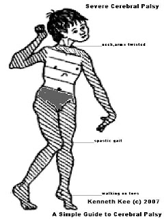
A Simple Guide to Cerebral Palsy


A Simple Guide to Cerebral Palsy
---------------------------------------

What is Cerebral Palsy?
-----------------------------

Cerebral palsy refers to a group of disorders characterised by loss of motor functions or any other nerve functions that appear in infancy or early childhood and permanently affect body movement and muscle coordination but don't worsen over time.
Even though cerebral palsy affects muscle movement, it isn't caused by problems in the muscles or nerves. These disorders are caused by brain damage that occurs during foetal development or near the time of birth.

What are the causes of Cerebral Palsy?
---------------------------------------------

The majority of children with cerebral palsy are born with it, although it may not be detected until months or years later.
Cerebral Palsy is mainly caused by brain damage that occurs during foetal development.
A small number of children have cerebral palsy as the result of brain damage in the first few months or years of life:
1. brain infections such as bacterial meningitis or viral encephalitis
2. head injury from a motor vehicle accident, a fall, or child abuse.

It is not an inherited disease.

What are the Symptoms of Cerebral Palsy?
---------------------------------------------------

The early signs of cerebral palsy usually appear before a child reaches 3 years of age. Babies with Cerebral Palsy are usually slow to reach developmental milestones.
Abilities like learning to roll over, sit, crawl, smile or walk are not well developed.

The motor abilities will often seem unusual and strange.
The most common are:
1.lack of muscle coordination when performing voluntary movements (ataxia);
2.stiff or tight muscles and exaggerated reflexes (spasticity);
3.walking with one foot or leg dragging;
4.walking on the toes,
5.crouched gait, or a "scissored" gait:
6.muscle tone that is either too stiff or too floppy.

The symptoms of Cerebral Palsy vary from just mild clumsiness to extensive uncontrolled muscle spasticity.
Many with cerebral palsy have other disabilities :
1.seizures,
2.an inability to see, hear, speak or learn as others do,
3.psychological or behavioural problems.

Not everyone with Cerebral Palsy is retarded. Some are of average intelligence.
While some people with Cerebral Palsy have learning disabilities many others do not.

What are the Types of Cerebral Palsy?
---------------------------------------------

There are four types of Cerebral Palsy:
1.Spastic Cerebral Palsy, the most common type, is a disorder in which certain muscles are stiff and weak.
2.Athetoid (dyskinetic, hypotonic, dystonia) Cerebral Palsy where involuntary movements are present.
3.Ataxic Cerebral Palsy occurs when the Cerebellum has been damaged, thus causing lack of coordination and jerky movements. This form of Cerebral Palsy have staggered or fragmented movements often involving tremors or exaggerated posturing (athetosis) and bizarre twisting motions.
4.Mixed Cerebral Palsy is when two or more types of cerebral palsy are present in the same person.

What is the Treatment of Cerebral Palsy?
-------------------------------------------------

Cerebral Palsy is a lifelong condition that cannot be cured.
The treatment is aimed at maintaining or improving the quality of life, providing the sufferer with tools to enjoy a near-normal life.

There are basically three types of treatment for Cerebral Palsy:
1.Surgery can be used to correct muscle contractures - removal of tight contractures that inhibits proper movement of the limbs.
2.Drugs such as clonazepam, baclofen and dantrolene are sometimes used to control muscle spasticity. Anticholinergic medications can help to control abnormal movements.
Alcohol or botulinum toxin type A (Botox) injections into muscle may be used to reduce spasticity for a short time so health care providers can work to lengthen a muscle.
3.Adjunctive therapies include
a.Physiotherapy, the most common treatment for cerebral palsy, consists of special exercises designed to increase and improve the range of movement and strength of the muscle groups, is aimed at strengthening and stretching muscles and preventing spasticity as well as increasing muscle control.
b.Occupational therapy is designed to help the child develop the fine motor skills needed to function day-to-day at home and school.
c.Speech therapy will also help the child develop communication skills.

What is the prognosis of Cerebral Palsy?
-------------------------------------------------

Cerebral palsy doesn't always cause profound disabilities.
A child with severe cerebral palsy might be unable to walk and need extensive, lifelong care.
Another with mild cerebral palsy might be only slightly awkward and require no special assistance.

Supportive treatments, medications, and surgery can help many individuals improve their motor skills and ability to communicate with the world.

Subscribe Now: Feed Icon

Clicktale

Click and bookmark these Social Networking Bookmarks

Social Bookmarking bookmark at folkd

Labels

Is the medical Guide simple enough?

Ads by Adbrite